Platycephalidae: M II A3
Unknown
Egg diameter in µm |
Number of oil globules |
Diameter of oil globule in µm |
Yolk texture |
Perivitelline space |
Position of oil globule at hatch |
Gut length at eye- pigment stage |
Myomeres |
1100-1225 |
multiple |
NA |
clear |
narrow |
astern |
48% of NL |
26 |
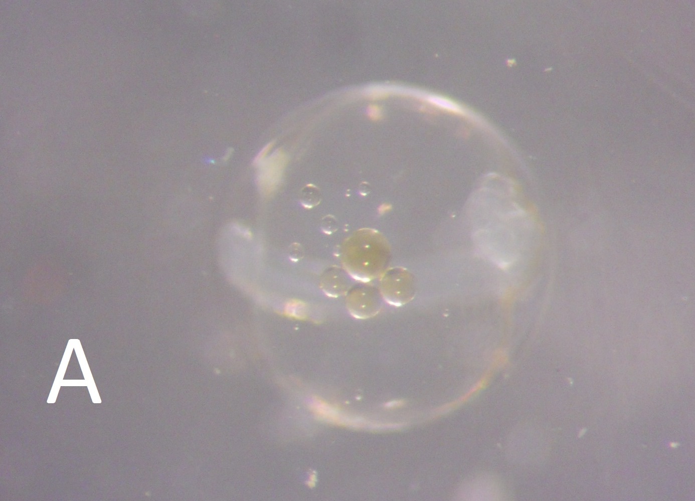
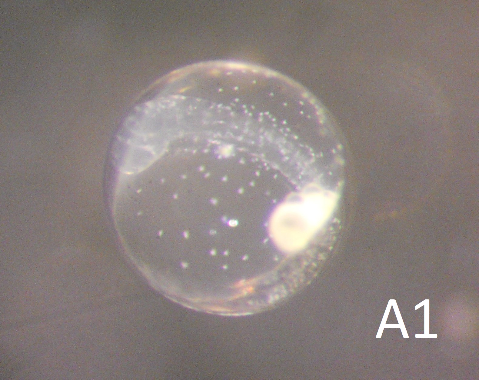
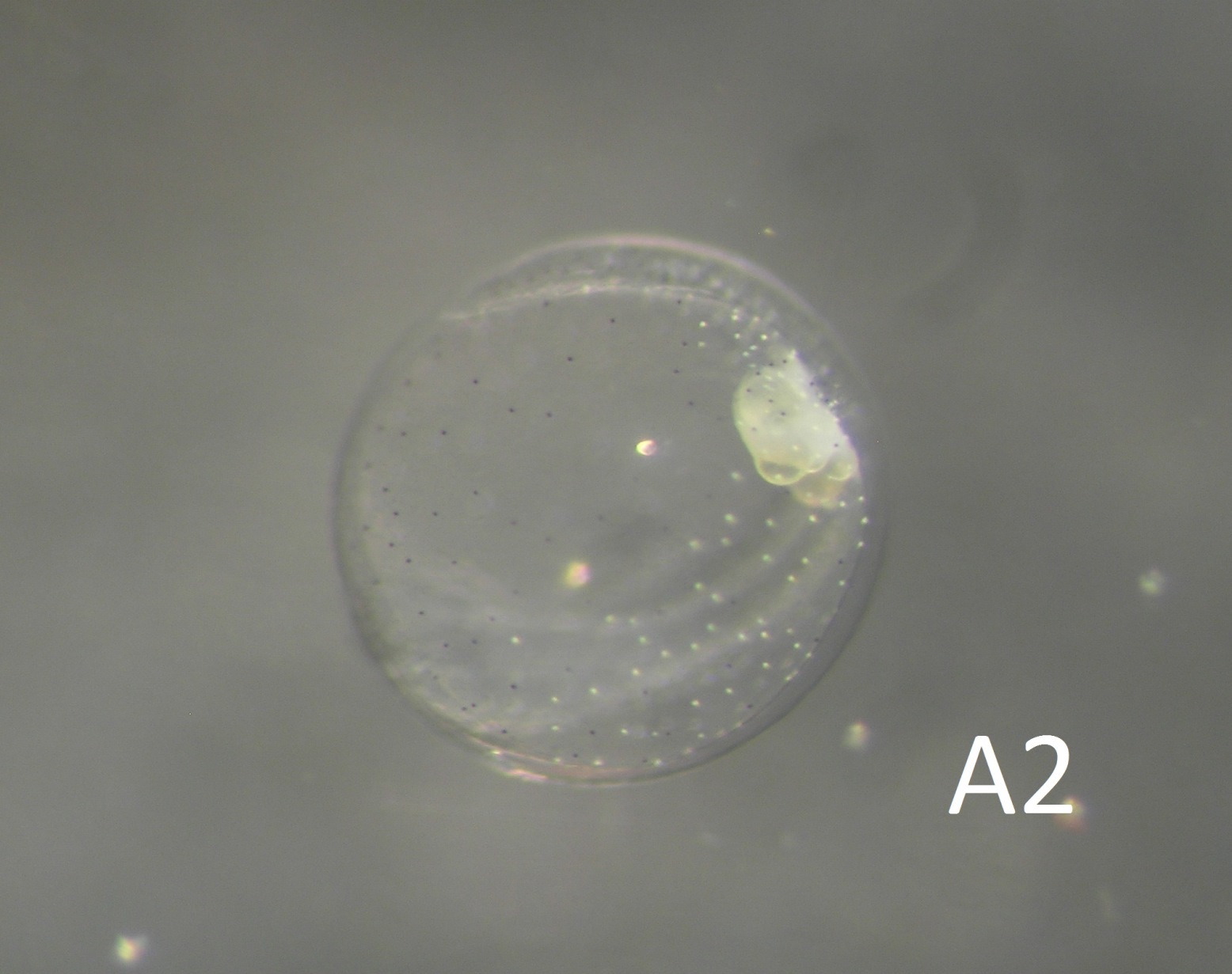
Egg: The oil globules are light amber (A), and clustered together, later in a white haze as the egg develops (A1, A2). White chromatophores appear on the yolk surface, interspersed with black spots. White chromatophores also cluster on the developing larva (A1). The yolk was noted as milky.
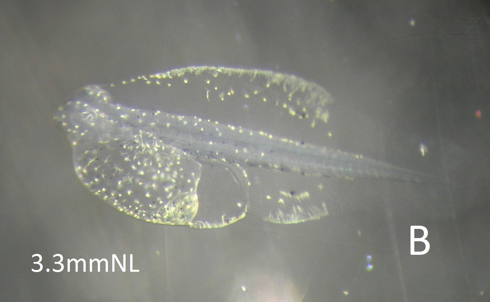
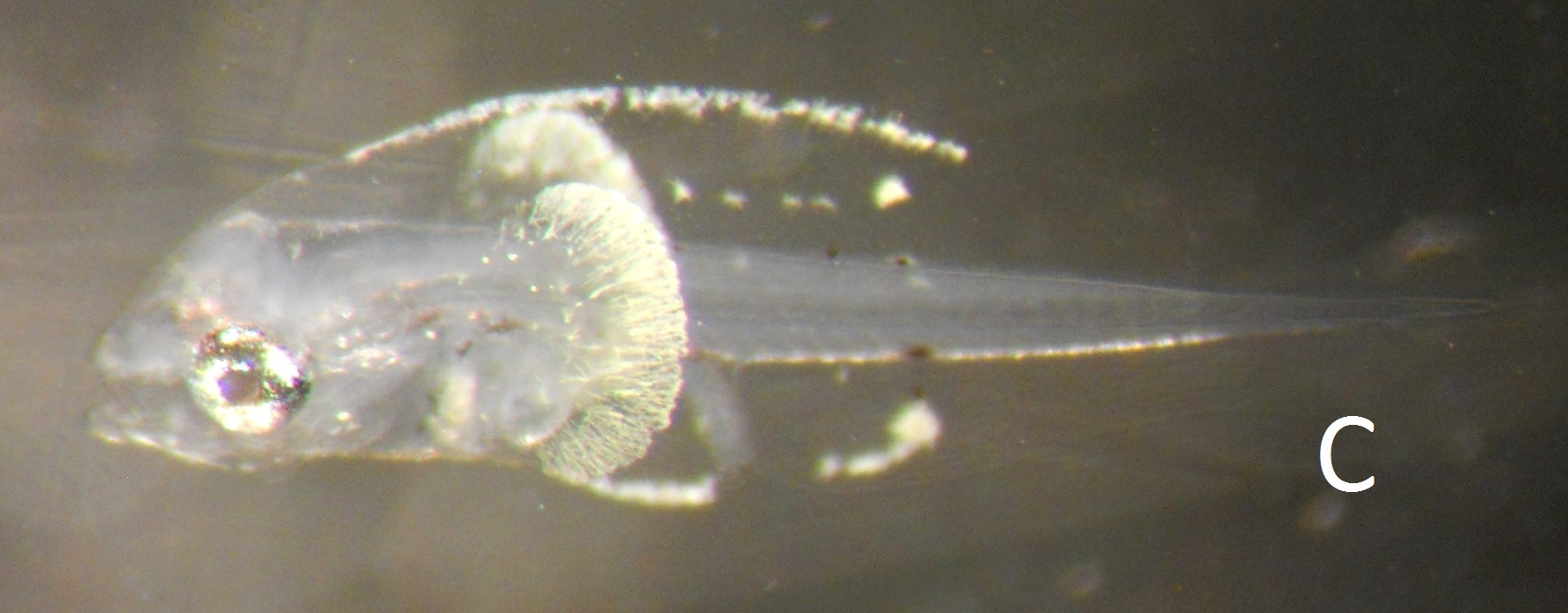
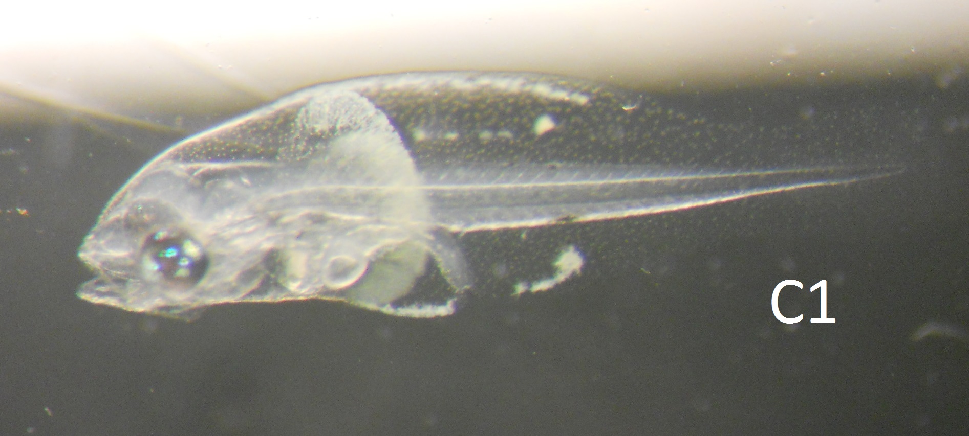
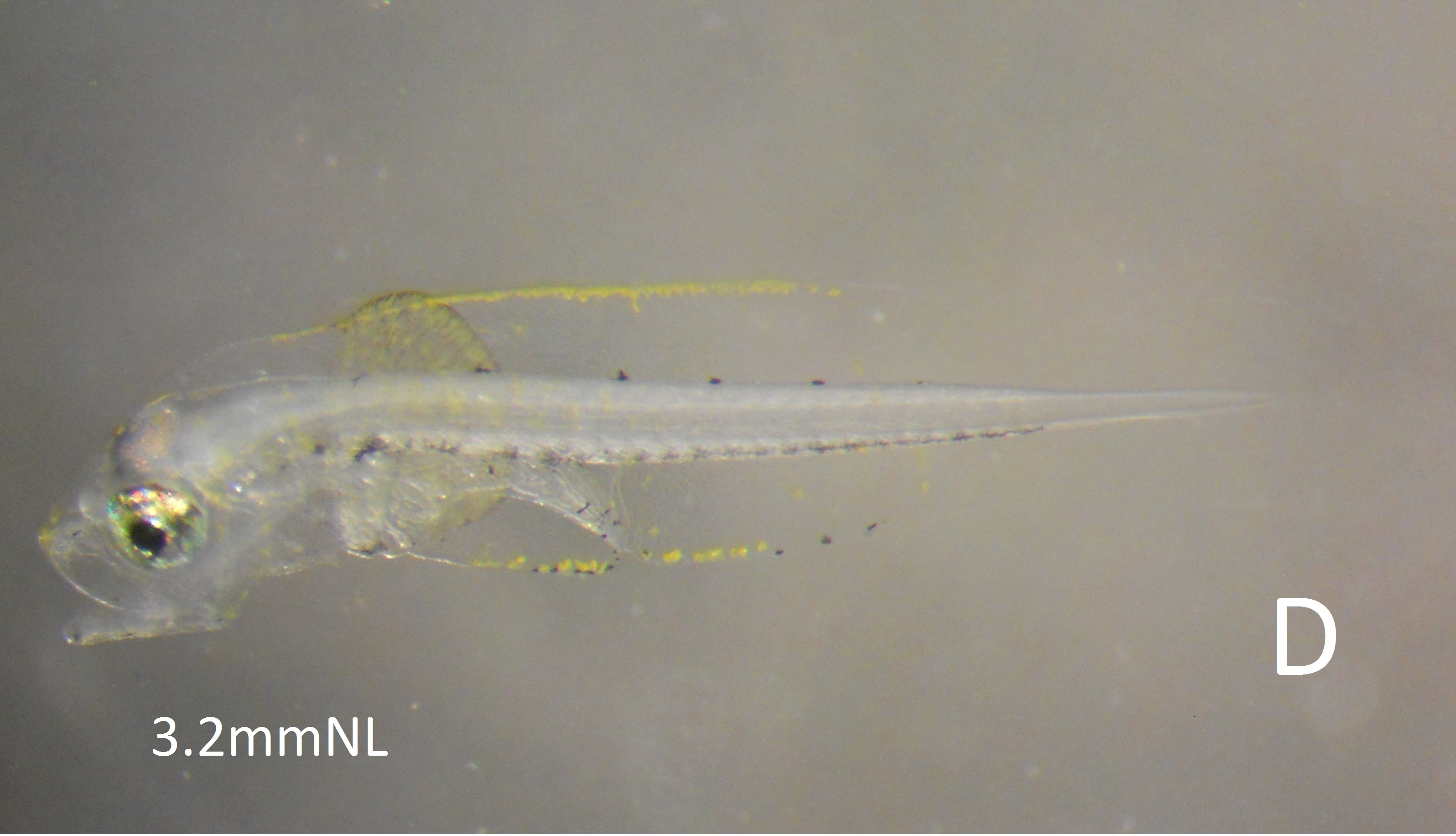
Larva: The yolksac of the early larva is covered in yellow spots, while those in the finfold edges are more smudgy and green (B). The finfolds of the 2-day larva have a distinctly "goose-pimpled" surface. Black spots are apparent on the dorsal and ventral edges of the notochord from day 1, and can even be seen on the lower finfold edge at day 4 (D).
This egg is easily confused with Parachirus xenicus (MIIA2A), but the white chromatophore pattern on the newly hatched larva of Parachirus is distinctive. An egg collected in the harbour entrance in November 2005 was probably this species, based on a photo taken at the time, although listed as Parachirus. The egg has only been seen on 3 occasions off Park Rynie, in February 2009, March 2012 and Auguat 2013, on each occasion as single eggs. The former two have been barcoded, and are closely associated with the platycephalid Papilloculiceps longiceps, in my tree, though about 8% different (BOLD).